Practical tip
Reliable diagnosis of approximal caries: How to optimise bitewing exposures?
In contrast to imaging techniques that aim to fully visualise the tooth roots, bitewing radiographs focus on other anatomical structures. In terms of caries diagnostics, the focus of the intraoral X-ray is on checking the jawbone and the interdental spaces.
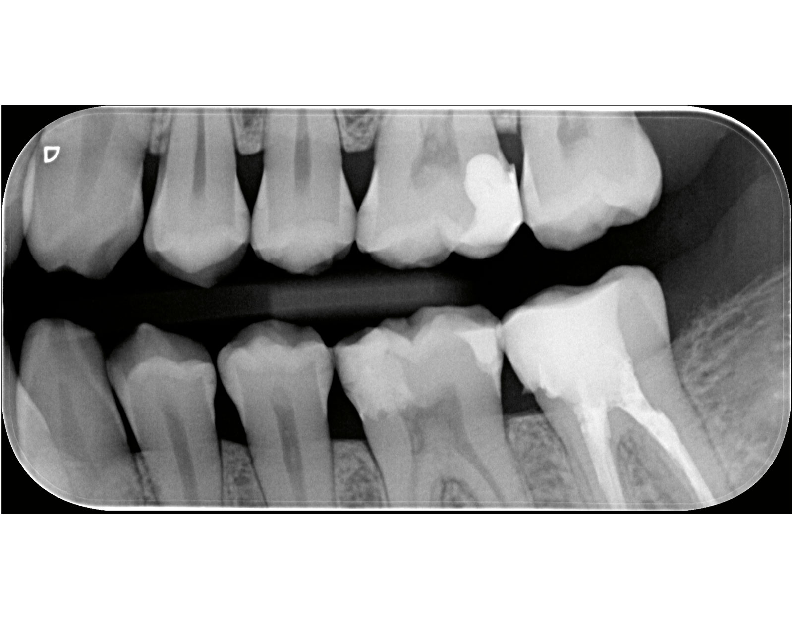
This is what matters: A bitewing image shows the posterior teeth in the upper and lower jaw on one half of the face.
If the dental assistants use the VistaPosition PSP holder system, they position the imaging plate between the tongue and dental arch of the side to be exposed and actively guide the bite plate downwards until contact is made with the occlusion.
The tube is then aligned parallel to the indicator arm on the aiming ring on the patient's cheek. Markings on the aiming ring help to adjust the collimator according to the position of the imaging plate.
Challenges:
- The dental hygienist usually places the imaging plate for the bitewing acquisition between the tongue and the dental arch. If it is positioned too close to the dental arch, the plate can press against the palate and the patient cannot close their mouth. As a result, the imaging plate is not positioned securely in place and the distance between the upper and lower jaw is too large
The interdental spaces are shown superimposed
- The premolars are not fully visualised
Tips from the practice:
- If the patient is unable to close the mouth after placement of the imaging plate due to a high oral floor or a tapered palatal plate, it is helpful to position the imaging plate on the back of the tongue. There is more space in the centre of the oral cavity.
To prevent the interdental spaces from overlapping, the central beam must be aligned orthoradially, i.e. tangentially to the selected contact surface. In simple terms, this means that it glides through the interdental spaces without touching them
- If the premolars are not completely imaged, the dental hygienist also places the image plate on the back of the tongue. If a holder from the VistaPosition PSP kit is used, the user rotates it so that the plate is aligned parallel to the premolars. This can be easily realised by positioning it on the back of the tongue. The front edge of the imaging plate should be positioned as close as possible behind the anterior region
See for yourself