Imaging
VistaVox S – Field of view: See what you want to see
Field of view
VistaVox S offers an ideal 3D volume that is adapted to the jaw arch. The special feature of VistaVox S is that its imaging volume is based on the human anatomy, representing precisely the area you need for diagnostics in the dental region. See what you want to see.
See more thanks to larger field of view
The jaw-shaped field of view of the VistaVox S maps the diagnostically relevant range of a 130-mm volume and is therefore visibly larger than the most commonly used volume of Ø 80 x 80 mm. The advantage is that thanks to this optimised volume shape, VistaVox S also completely covers the region of the rear molars.

Additional volumes ø 50 x 50 mm in 120 µm or 80 µm voxel size
In addition to jaw-shaped images, VistaVox S offers ten further Ø 50 x 50 mm volumes: five each for the upper jaw and for the lower jaw. These are used if the indication only requires imaging of a certain region of the jaw, e.g. for endodontical or implantological treatments. Depending on the required level of detail in the image, the volumes can be used with a voxel size of either 80 or 120 µm.

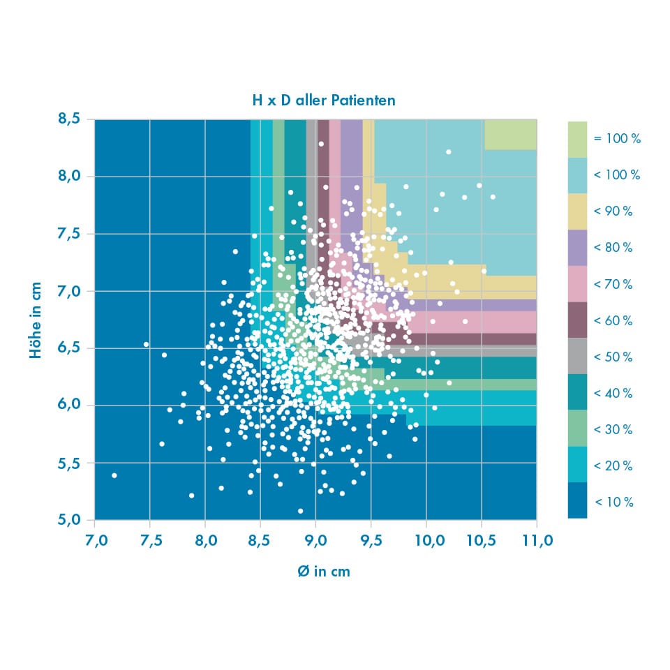
Investigations on the required field of view for 3D diagnostic imaging in dentistry
1,020 patients were examined in a study from Dr Johannes Krause. The study shows that a volume with a height of 85 mm and diameter of 110 mm is required for 100% coverage of the dental region. With a volume of Ø 80 x 80 mm, only around 1.4% of all patients can be covered in full. By contrast, the adapted, jaw-shaped volume of the VistaVox S covers the dental region of all patients.*